OtoLens is the outcome of a research project aimed at leveraging artificial intelligence and image processing to analyze otoscope images. Developed with the primary goal of educating users about AI applications in healthcare, OtoLens allows users to capture and analyze images of the ear using their mobile device’s camera or photo album. This application is intended for educational purposes only and should not be used as a diagnostic tool. This app is currently in the model research and development phase, and errors may sometimes occur. Once we complete field testing and fix all errors, we will deliver the final version to our application delivery team. For detailed information on the latest features and updates, please refer to the release notes in the Changes section.
Research Team:
1.
2.
3.
4.
Key Functionals:
Image Quality Assessment: The app includes a preliminary classifier that evaluates the quality of the selected image. It ensures that only images meeting a certain quality threshold are processed further, enhancing the accuracy of the analysis.
OME (Otitis Media with Effusion) Classifier: Identifies signs of OME, a condition characterized by fluid in the middle ear without symptoms of acute infection.
TMP (Tympanic Membrane Perforation) Classifier: Detects perforations or holes in the tympanic membrane (eardrum).
Myringitis Classifier: Detects inflammation of the tympanic membrane, often associated with redness and irritation.
Installation:
Download and Install TestFlight:
- Go to the App Store and search for “TestFlight.”

- Download and install the TestFlight app on your device.
Request Invitation:
- Send an email to project admin requesting access to the OtoLens app.
Receive Invitation:
- You will receive an invitation link via email. Open the email on your device.
Join the Beta:
- Click the invitation link in the email, which will open TestFlight and prompt you to join the OtoLens beta.
Install OtoLens:
- Follow the instructions in TestFlight to download and install the OtoLens app.
Open OtoLens:
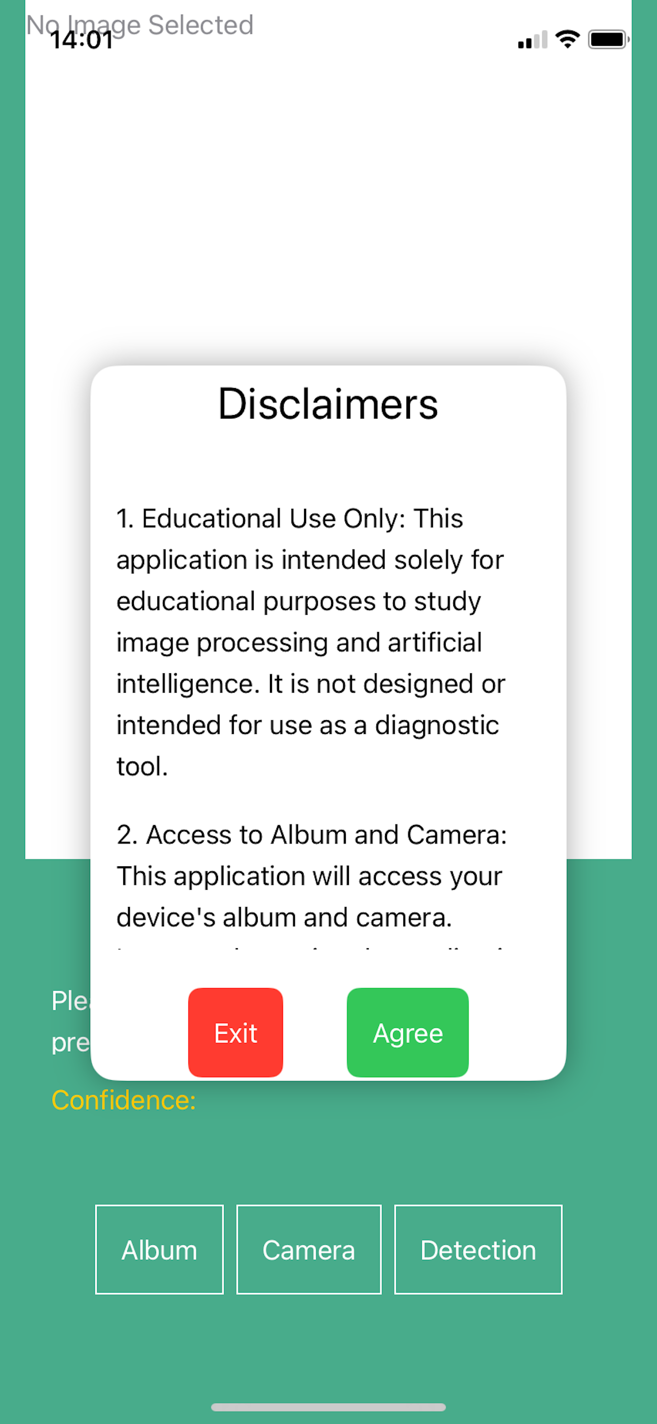
- Once installed, open the OtoLens application and grant the necessary permissions for accessing your camera and photo album.
Using the Album:
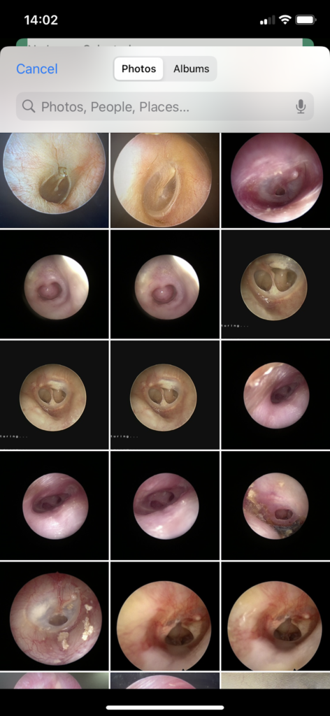
1. Tap the “Album” button to select an image from your device’s photo library.
2. Choose an otoscope image for analysis.
3. The app will automatically assess the image quality and proceed with the analysis if the quality is acceptable.
Using the Camera:
1. Tap the “Camera” button to capture a new image using your device’s camera.
2. Ensure the image is clear and focused for accurate analysis.
3. The app will assess the quality of the captured image and proceed with the analysis if the quality is acceptable.
Interpreting Results:
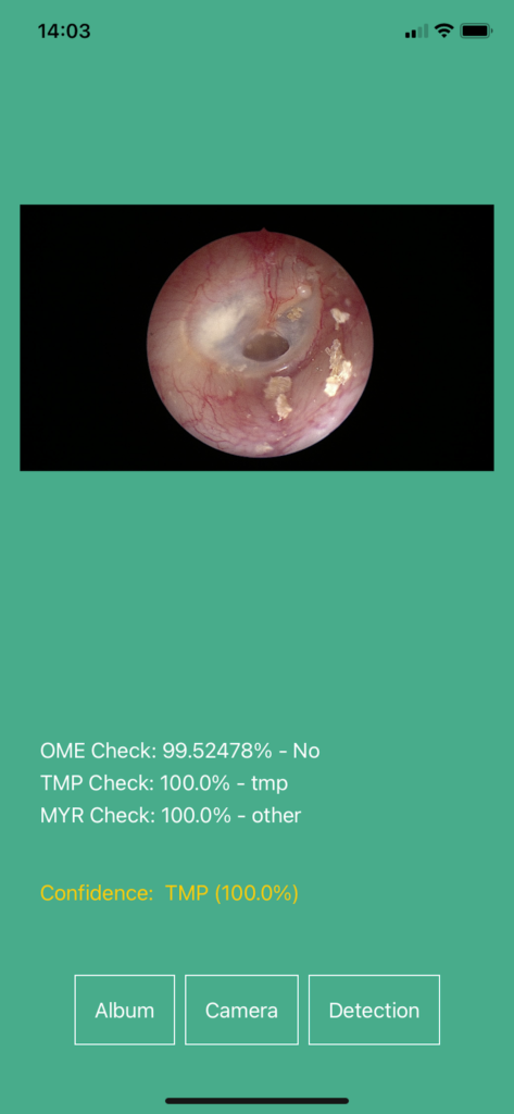
1. Tap the “Detection” button to initiate the analysis process.
2. The app will display the results, including the likelihood of various ear conditions such as Otitis Media with Effusion (OME), Tympanic Membrane Perforation (TMP), and Myringitis.
3. The results will be presented with the confidence levels of the AI model’s predictions. Note that the application will also indicate if the image quality is not suitable for analysis.
How to Interpret Classifier Results
When using OtoLens, the results from the classifiers will provide insights into the detected conditions of the tympanic membrane and middle ear. Here’s a guide on how to interpret these results effectively:
- Understanding the Output
- Each classifier (OME, TMP, MYR) will produce a result indicating one of three categories: “normal,” the specific symptom (OME, TMP, or MYR), or “other.”
- Alongside the category, a confidence percentage will be provided, indicating the model’s confidence in its classification.
- Result Categories and Actions
- Normal: If all classifiers indicate “normal” with a confidence level of 80% or higher, the result is considered normal. This suggests no significant pathology was detected in the images.
- Other: If all classifiers indicate “other” with a confidence level of 80% or higher, the result is considered as “other.” This indicates that the images do not fit the patterns associated with the specific symptoms (OME, TMP, MYR) and may require further investigation.
- Symptom (OME, TMP, MYR): If any classifier detects a symptom (OME, TMP, or MYR) with a confidence level of 80% or higher, the result will highlight the symptom in descending order of confidence. This indicates a high likelihood of the detected condition and should be further examined by a healthcare professional.
- Mixed Results Interpretation
- High Confidence Symptoms: If any classifier reports a symptom with high confidence (80% or higher), the recommendation will focus on these symptoms, ignoring “normal” and “other” classifications.
- No High Confidence Symptoms: If no classifiers report a symptom with high confidence, but there are “normal” or “other” classifications with confidence levels above 80%, the recommendation will reflect the most confident result among these.
- Low Confidence Across All: If all classifiers report confidence levels below 80%, the result will be categorized as “Out of model,” indicating that the image data does not clearly fit any of the trained models.
- Example Interpretations
- Example 1:
- OME Check: 94.71% Normal
- TMP Check: 99.99% Normal
- MYR Check: 100% Other
- Interpretation: Since there is no symptom with confidence >= 80%, but “Normal” and “Other” categories have high confidence, the result will prioritize the most confident “Other” category.
- Example 2:
- OME Check: 97% Normal
- TMP Check: 99% Other
- MYR Check: 100% Other
- Interpretation: The result will prioritize “Other” due to high confidence in “Other” categories.
- Example 1:
- Limitations
- Data and Model Constraints: The model’s current version is based on a limited dataset. As such, results may vary, and continuous feedback and retraining are essential for improving accuracy.
- Non-Diagnostic: Remember, the app is for educational purposes only and is not a diagnostic tool. Always seek professional medical advice for health-related decisions.
For detailed information on the latest features and updates, please refer to the release notes in the Changes section.
Disclaimer
1. Educational Use Only: This application is intended solely for educational purposes to study image processing and artificial intelligence. It is not designed or intended for use as a diagnostic tool.
2. Access to Album and Camera: This application will access your device’s album and camera. Images taken using the application will be saved to your device’s album.
3. Data Collection: This application does not collect, store, or transmit any user data beyond the images processed within the app.
4. Limitation of Liability: The author of this application shall not be held liable for any outcomes or consequences resulting from the use of this application. The recommendations provided by the application are for educational purposes only and should not be construed as medical advice.
5. Model Limitations: This app is designed for use with images from an otoscope only. The model is built from a very limited data set. The data you take may be out of the model scope we trained.
Technology Behind OtoLens
OtoLens utilizes advanced AI and machine learning technologies to analyze otoscope images. Key technologies include:
CoreML: Apple’s framework for integrating machine learning models into iOS applications. CoreML enables OtoLens to perform fast and efficient on-device image analysis.
CreateML: Apple’s tool for training custom machine learning models. The OtoLens models were trained using CreateML with a curated dataset of otoscope images.
Vision Framework: Used for handling image processing tasks such as image quality assessment and feature extraction.
Swift: The application is built using Swift, Apple’s powerful and intuitive programming language for iOS development.
Current Model Limitations
Potential Misclassification of Tympanic Membrane Perforation (TMP)
One key challenge with the current model is the potential for misclassification when dealing with tympanic membrane perforations. The presence of a hole or a shape resembling a hole in the eardrum can lead to the model incorrectly identifying the condition as TMP, especially if no more intense symptoms are present to suggest an alternative diagnosis like Myringitis (MYR).
Dependency on Image Quality for OME and MYR Confidence
The accuracy of Otitis Media with Effusion (OME) and Myringitis (MYR) classifications heavily relies on the quality of the images. Suboptimal lighting, focus, or angles can significantly reduce the confidence levels of these classifications, leading to potential misdiagnosis or the need for re-evaluation with better-quality images. Rational Behind Model and Preprocessing Design
Middle Ear Pathology Image Analysis: Symptoms and Characteristics
Normal Eardrum
A normal eardrum typically appears pearly grey or light pink, with a translucent and slightly shiny surface. Key features include a visible light reflex (“cone of light”) in the anterior inferior quadrant and landmarks like the malleus handle. The membrane should be neutral, showing no signs of bulging, retraction, or perforation and no visible fluid or air bubbles behind it.
Otitis Media with Effusion (OME)
OME is characterized by a dull or opaque tympanic membrane, often with a yellow, amber, or blue tint due to fluid behind the membrane. The light reflex is usually decreased or absent, and the membrane may be retracted or bulging. Visible air-fluid levels or bubbles and prominent blood vessels due to inflammation indicate fluid accumulation in the middle ear space without acute infection.
Tympanic Membrane Perforation (TMP)
TMP is marked by a visible hole or tear in the eardrum, with irregular edges and possible discharge if infection is present. The light reflex is altered or absent, and the perforation may make middle ear structures visible. The size and location of the perforation can vary significantly, resulting from trauma, infection, or chronic eustachian tube dysfunction.
Myringitis
Myringitis manifests as tympanic membrane inflammation, with redness, irritation, and sometimes tiny blisters or vesicles. This inflammation can lead to thickening or clouding of the membrane, altering its transparency and light reflex. In bullous myringitis, more prominent fluid-filled blisters may be present. Symptoms include ear pain and hearing loss, often due to viral or bacterial infections. Image Analysis Approach.
To differentiate between normal and pathological conditions, careful examination of various features is essential:
Color and Transparency: Assessing the eardrum’s color and translucency.
- Light Reflex: Evaluating the presence and quality of the light reflex.
- Membrane Position: Observing for bulging, retraction, or neutral position.
- Visible Structures: Identifying any structures behind the membrane.
- Membrane Integrity: Checking for perforations or abnormal growths.
- Inflammation Signs: Noting redness, irritation, or blister presence.
By systematically analyzing these characteristics, clinicians and automated systems can make more accurate diagnoses and ensure appropriate treatment plans are developed. However, it is crucial to acknowledge the model’s limitations and continually refine its capabilities to enhance diagnostic accuracy and reliability.
Changes Section
OtoLens Release Notes – Version 1.0
Features
– Image Capture: Capture images using your mobile device’s camera or select images from your photo album.
– Image Quality Assessment: Automatically assess the quality of the captured image to ensure it meets the required standards for analysis.
– Image Preprocessing: Enhance images through preprocessing techniques to improve the accuracy of subsequent analysis.
Classifier Features
– OME Classifier: Detects signs of Otitis Media with Effusion. Very limited in the first version – need feedback to improve.
– TMP Classifier: Identifies Tympanic Membrane Perforations. Very limited in the first version – need feedback to improve.
– Myringitis Classifier: Recognizes signs of Myringitis. Very limited in the first version – need feedback to improve.